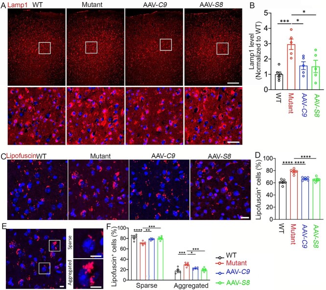
Figure 4.
C9orf72 and Smcr8 rescued lysosomal abnormalities in C9FTD/ALS mutant mice. (A) Representative confocal image of motor cortex from 20-month-old mice stained with antibodies against Lamp1 (red) and Hoechst stains for nuclei (blue). The lower panels are enlargements of white boxed areas in the upper panels. Scale bars: 200 μm (upper panels), 20 μm (lower panels). (B) Quantification of Lamp1 staining signal intensity normalized to WT. Note that AAV-C9orf72 or AAV-Smcr8 expression rescued the aberrantly increased Lamp1 intensity in C9TFD/ALS mutant mice. (C, E) Representative confocal images of lipofuscin accumulation in a 20-month-old mouse motor cortex. Hoechst stains nuclei (blue). Right panels in E are enlargements of white boxed areas in the left panels. Scale bars: 20 μm in C and 10 μm in E right panels. (D) Quantification of the percentage of lipofuscin-positive cells. (F) Quantification of the percentage of sparse and aggregated lipofuscin-positive cells out of total lipofuscin-positive cells. Mutant: C9orf72+/−;C9-BAC; AAV-C9: C9orf72+/−;C9-BAC with AAV-PHP.eB-C9orf72; AAV-S8: C9orf72+/−;C9-BAC with AAV-PHP.eB-Smcr8. Data information: For all analyses, data are presented as mean ± SEM. N = 6 mice with one-way ANOVA with Bonferroni’s post hoc test (*P < 0.05, **P < 0.01, ***P < 0.001, ****P < 0.0001).

