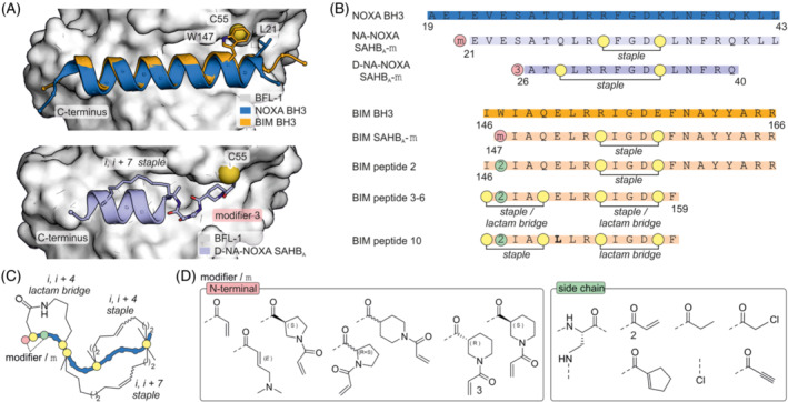
FIGURE 3.

(A) Top: Crystal structure of NOXA BH3 (blue, pdb: 3mqp) and BIM BH3 (orange, pdb: 2vm6) bound to BFL‐1 (pdb: 3mqp). Bottom: Crystal structure of i, i + 7 stapled covalent inhibitor D‐NA‐NOXA SAHB bound to C55 of BFL‐1 (pdb: 5whh). (B) Sequences and modification sites of BFL‐1, BCL‐2A1 and MCL‐1 binding peptides. (C) Scheme of the different employed crosslinks. (D) Chemical structures of tested modifiers.
