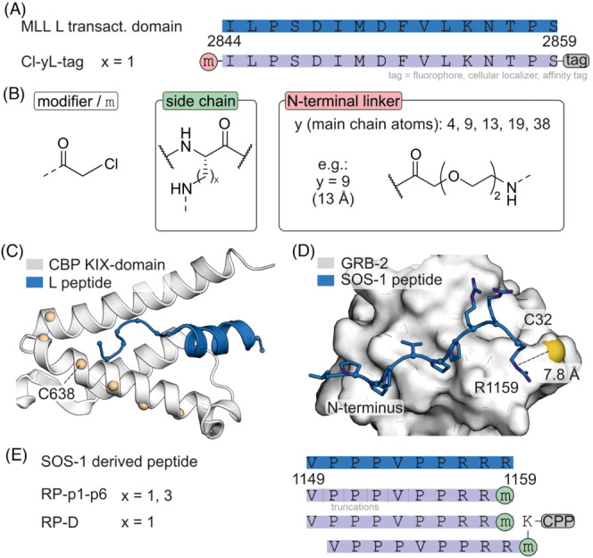
FIGURE 5.

(A) Sequence of MLL‐transactivation domain L and derived covalent binders with modification sites. (B) Chemical structures of modifiers (x = 1 or 3). (C) NMR structure (pdb: 2lxs) of KIX‐domain of CBP (white) and MLL‐transactivation domain (blue) with positions used for the generation of cysteine variants (beige). (D) NMR structure of GRB‐2 bound to a SOS‐1 derived peptide (pdb: 1gbq). (E) Sequence of SOS‐1‐derived peptide and derived monomeric and dimeric covalent inhibitors attached to a cell penetrating peptide (CPP).
