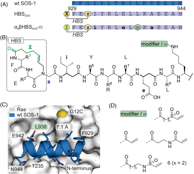
FIGURE 6.

(A) Sequence of wt SOS‐1 and derived inhibitors with modification sites (i: β‐isoleucine, e: β‐glutamic acid, a: β‐alanine, X: 4‐pentenoic acid, Z: 5‐hexenoic acid, z: N‐allylglycine). (B) Chemical structure of the hydrogen bond surrogate (HBS), selected β‐amino acids (bold), position of the modifier m and the HBS are shown. (C) Crystal structure of Ras (white, pdb: 1nvw) in complex with SOS derived peptide (wt SOS, blue, pdb: 1nvw, glycine was varied to cysteine for demonstration purposes). (D) Chemical structure of tested electrophiles (x = 1 or 2).
