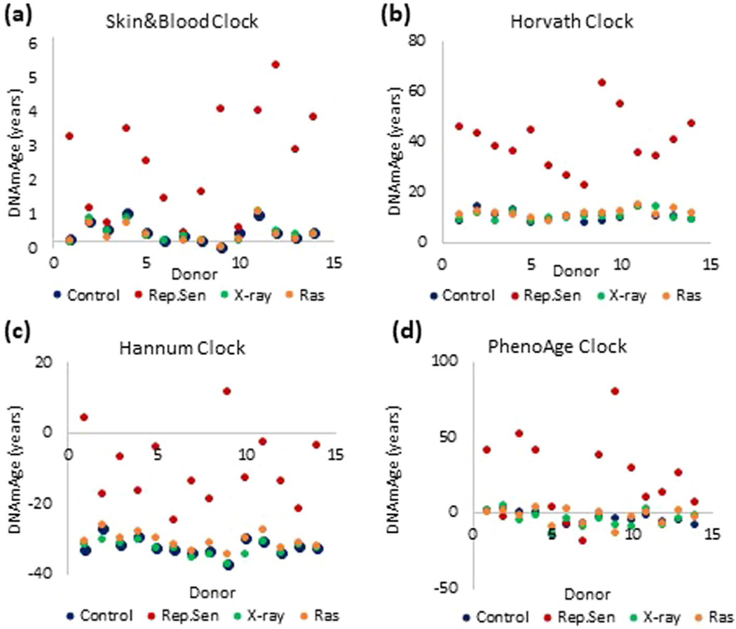
Extended Data Fig. 2 |. effects of various inducers of cellular senescence on epigenetic aging.
Measurement of EpiAge with four epigenetic clocks on primary human fibroblasts isolated from neonatal foreskins of 14 donors. Fibroblasts were cultured until replicative senescence (red), induced to senesce by X-irradiation (green), induced to senesce by ectopic expression of activated ras oncogene (orange) or untreated (blue). Methylation profiles of these cells were analyzed using the (a) Skin&blood clock, (b) Horvath clock, (c) Hannum clock and (d) PhenoAge clock.

