FIGURE 10.

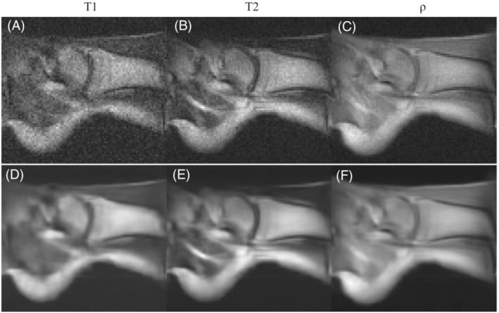
In vivo images of the right ankle of a healthy volunteer taken at i3M with 3D‐RARE sequences and reconstructed with FFT. A–C, A selected slice for the and ‐weighted acquisitions respectively. D–F, The same as A–C but BM4D filtered

In vivo images of the right ankle of a healthy volunteer taken at i3M with 3D‐RARE sequences and reconstructed with FFT. A–C, A selected slice for the and ‐weighted acquisitions respectively. D–F, The same as A–C but BM4D filtered