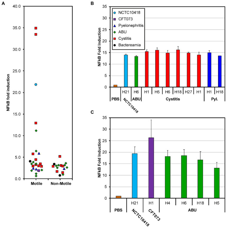
Figure 2.
NF-kB response of bladder RT4 cells challenged with whole cells and flagellin. (A) NF-κB fold induction of motile and non-motile E. coli clinical isolates [Supplementary Table S1 (3,398–3,710) and Supplementary Table S2 (3,399–3,707)]. Points have been scattered left or right with respect to the x-axis for clarity. Colors and point shape as shown in the key of panel (B) represent the associated type of infection of the isolate (B) NF-κB response of RT4 bladder cells challenged for 24 h with 250 ng/mL flagellin filaments isolated from 11 different E. coli strains; from left to right: NCTC10418, 3,419, 3,408, 3,409, 3,411, 3,412, 3,414, 3,417, 3,398, 3,424 (Supplementary Table S1). (C) NF-κB fold increase of RT4 bladder cells challenged with 50 ng/ml flagellin filaments isolated from 6 different E. coli strains; from left to right: NCTC10418, CFT073, 3,692, 3,693, 3,694, 3,698 (Supplementary Table S1). Strain choice in (B,C) reflects the diversity in whole cell challenge responses presented in (A). Error bars represent the standard deviation. All data is an average of a minimum of n = 3 independent biological repeats with n = 2 technical repeats for NF-κB assays.

