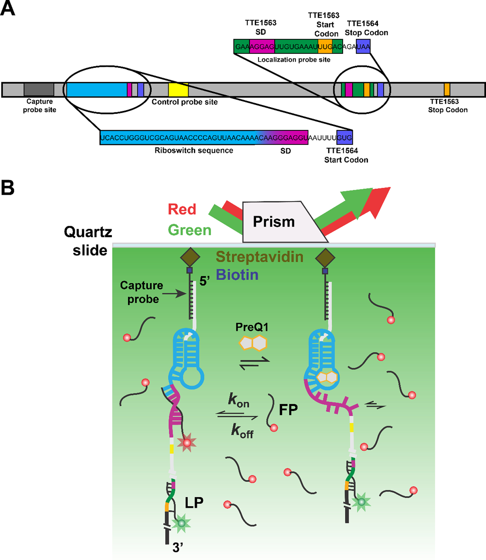
Fig. 1.

SiM-KARTS measurements of an mRNA with embedded preQ1 riboswitch. (A) The preQ1 riboswitch containing mRNA. The transcript is immobilized onto the microscope slide after hybridization of the biotinylated capture probe (CP) to the corresponding recognition site on the target. The distally hybridized localization probe (LP) allows for the localization of single immobilized mRNA molecules. Transiently binding fluorescence probe (FP, here against the Shine-Dalgarno (SD) sequence of the TTE1564 protein) senses structural changes of the riboswitch. (B) Experimental schematic of the SiM-KARTS assay using a prism-based TIRFM setup. After the RNA complex is immobilized onto a microscope slide via streptavidin and biotinylated BSA (biotin-BSA, not shown for simplicity), it is illuminated with a 532 nm wavelength laser (exciting the LP) to identify the RNA complex. A red laser (638 nm) is used to excite the FP, whose repeated binding to the Shine-Dalgarno (SD) sequence results in spikes of fluorescence. In the presence of preQ1, the accessibility of the binding site of the FP changes.
