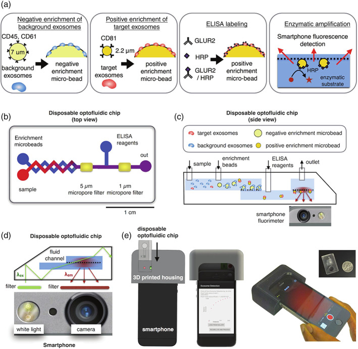
FIGURE 3.

Design of a microbead‐based mobile extracellular vesicle (EV) detector. (a) Schematic overview of microbead immunoassay procedure from EV enrichment to fluorescent signal emission. (b) Graphical representation of optofluidic chip, and (c) a side view of chip and phone interplay with bead assay. (d) Set‐up of LED and smartphone camera for excitation and emission. (e) Rendered image of 3D‐mount for smartphone and disposable chip integration. Reproduced (adapted) under creative commons license from Ko et al. (2016)h
