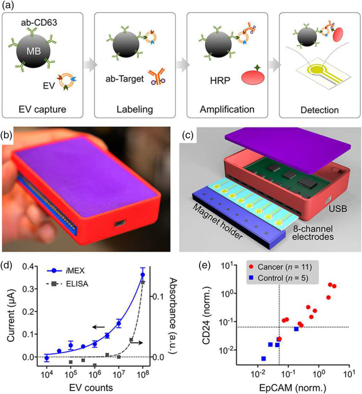
FIGURE 8.

Summary of iMEX approach. (a) Schematic representation of iMEX assay. CD63‐ positive EVs are captured on magnetic beads in plasma and labeled with HRP for electrochemical detection. (b) The iMEX device. (c) Eight electrode set‐up of the iMEX. Magnets below the electrodes concentrate immunomagnetically captured EVs. (d) iMEX and ELISA response comparison to titrated concentrations of EVs. (e) iMEX analysis of plasma samples from ovarian cancer patients and healthy controls. Adapted with permission from Jeong et al. (2016) and Shao et al. (2018). Copyright 2016 and 2018 American Chemical Society
