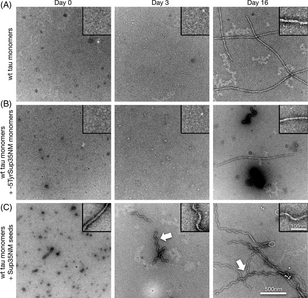
FIGURE 2.

Sup35NM seeds wild‐type tau aggregation in vitro. A‐C, Transmission electron microscope images of negatively stained preparations of human 2N4R wild‐type tau monomer aggregation under low heparin conditions (A), and after addition of ‐5TyrSup35NM monomers (B), or Sup35NM seeds (C), at days 0, 3, and 16. Note the occurrence of corkscrew‐shaped tau fibrils at days 3 and 16 upon seeding with Sup35NM fibrils (arrows, C). Scale bar for all images: 500 nm, for all magnified insets: 100 nm
