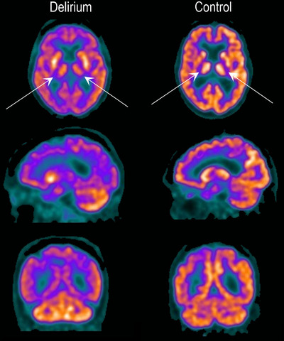
FIGURE 2.

18F‐fluorodeoxyglucose positron emission tomography (FDG‐PET) images. FDG‐PET during delirium compared to control. Top: axial; middle: sagittal; bottom: coronal slices. The delirium scan is an 89‐year‐old female with delirium but no dementia with acute gastroenteritis. The control scan is a cognitively intact 80‐year‐old male with pneumonia and acute kidney injury. Darker colors indicate lower metabolism. There is relative hypometabolism in the thalamus bilaterally (arrows) and also throughout the cerebral cortex in the delirious patient compared to cognitively intact control. During delirium, relative preservation of cerebellar FDG uptake is noted. See Appendix B in supporting information for further images
