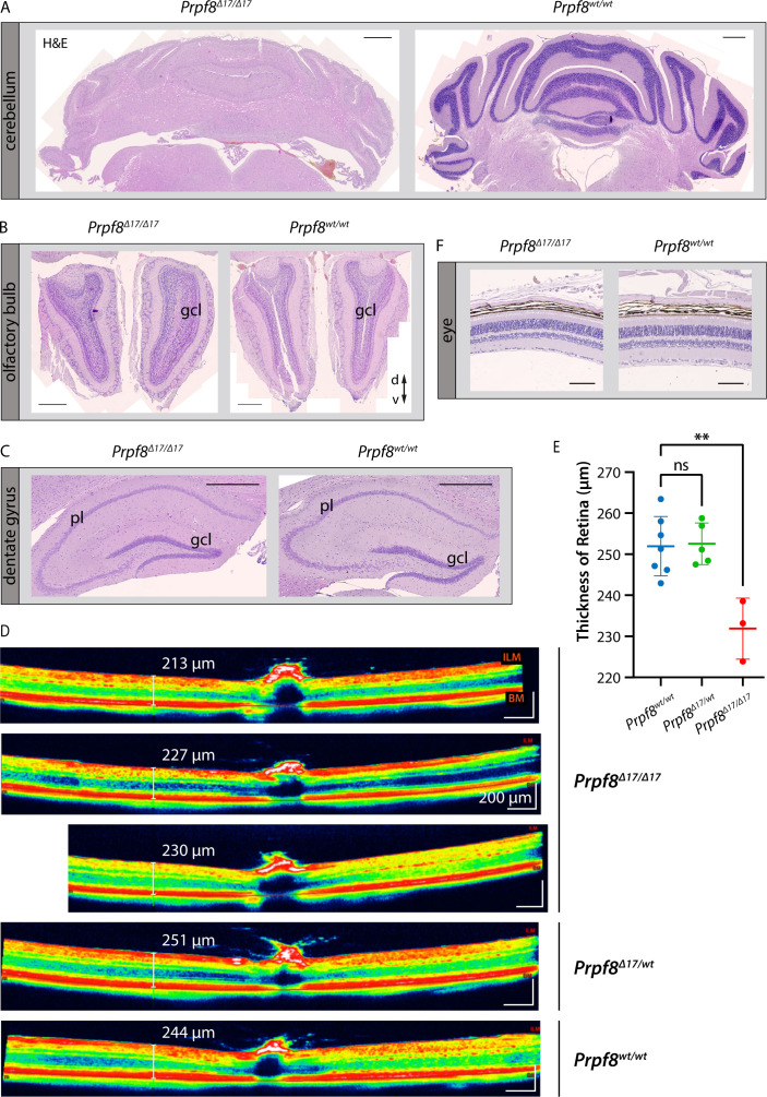
Figure S6. Histopathological inspection of condition of selected granule cell populations in the brain and eyes in the cohort of 22-wk-old mice of the Prpf8Δ17 strain.
(A, B, C) Although the cerebellar granule cell layer (gcl) was severely decreased (A), granular layers in the olfactory bulb (B), and in the dentate gyrus in hippocampus (C) did not show any signs of reduction when compared to the corresponding structure in a wt littermate. H&E, hematoxylin and eosin; gcl, granule cell layer; pl, pyramidal layer; d, dorsal; v, ventral. Scale bar = 500 μm. (D) Representative view of a cross-sectional image of retina gained by OCT examination. Retinal boundaries are indicated by red lines: ILM, internal limiting membrane (the boundary between the retina and the vitreous body); BM, Bruch’s membrane (the innermost layer of the choroid). (E) Evaluation of the retinal thickness in 22-wk-old cohorts of Prpf8Δ17 animals revealed a decrease in retinal breadth in homozygous mice (one-way ANOVA followed by Dunnett’s multiple comparison, P = 0.0017, **), whereas no difference was observed between heterozygous and control mice (ns). n = 7 for Prpf8wt/wt animals, n = 5 for Prpf8Δ17/wt, and n = 3 for Prpf8Δ17/Δ17 mice; all genotype conditions included animals of both sexes. (F) Representative image displaying preserved retinal layers in a homozygous Prpf8Δ17/Δ17 mouse and a control wt littermate. Scale bar = 100 μm.

