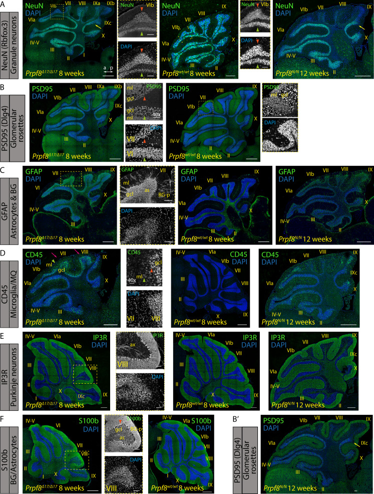
Figure S9. Immunohistochemical staining for markers of selected cerebellar cell types in 8-wk-old Prpf8Δ17/Δ17 and 12-wk-old Prpf8Y2334N/Y2334N mice.
(A) Rbfox3 (NeuN) immunolabeling was used to visualize nuclei of granule neurons. The detail shows an apoptotic zone in apical portion of lobule VIb (orange arrowhead) in contrast to its less affected vermian portion (green arrowhead). The boundary between posterior lobule IXb and lobule IVc indicated by the yellow arrow. a, anterior; p, posterior. (B,B′) Punctate pattern of PSD95 marks glomerular rosette in the granular layer and synaptic junctions of Purkinje dendrites. Detail: observed decay of the glomeruli (orange arrowhead); the PSD95 signal within the molecular cell layer (ml) was preserved. A retained, vermian portion of lobule VI is shown for comparison (green arrowhead). The boundary between posterior lobule IXb and lobule IVc indicated by the yellow arrow. ml, molecular layer; gcl, granule cell layer. Insets from Prpf8wt/wt animals are derived from area marked by yellow rectangles and magnified 2.5×. (C) Signal of GFAP was enhanced in the gcl of homozygous animals, confirming activation of astrocytes (as). The signal in the molecular layer (ml) represents GFAP-positive processes of Bergmann glia (BG-p). (D) CD45 immunostaining visualized phagocytic microglia and/or macrophages in the gcl of the degenerating posterior lobe lobules (pink arrows). The labeled microglia showed amoeboid morphology with thick cellular processes characteristic of DAMs (orange arrowhead). The signal was also present in the molecular layer (ml), which might coincide with clearance of residual parallel fibers from degenerated granule neurons (green arrowheads). (E) Purkinje cells were visualized based on the expression of IP3R that is present both in the somatodendritic compartment and in the axonal projections (ax). No major perturbance of Purkinje neurons was spotted even in the apoptotic areas (detail of lobule VIII). (F) Cell bodies of Bergmann glial cells were normally localized around Purkinje cell somata (orange arrowhead) and their radial processes (BG-p) properly terminated with endfeet at the pial surface, as revealed by staining with anti-S100 protein. The anti-S100b antibody in parallel visualized astrocytes (as) in the gcl. (A, B, C, D, E, F) Confocal immunofluorescence images of 5-μm FFPE sagittal cerebellar slices; scale bar = 200 μm; in insets, scale bar = 50 μm. Only the Prpf8wt/wt littermates of Prpf8Δ17/Δ17 animals are shown for clarity.

