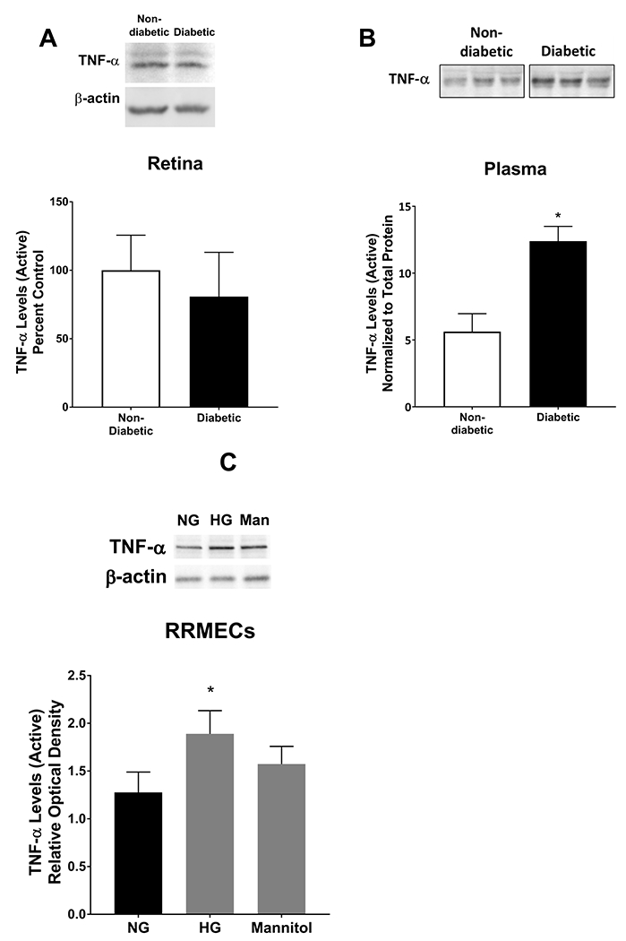
Figure 1. TNF-α levels under hyperglycemic conditions.

TNF-α levels in retinas, plasma, and RRMECs were determined using western blotting techniques. A) TNF-α levels in retinas collected from diabetic rats were not altered when compared to retinas collected from non-diabetic rats. However, there was a significant increase in TNF-α levels in plasma (B) collected from diabetic rats when compared to controls. C) TNF-α in RRMECs grown under hyperglycemic (HG) conditions had significantly higher levels when compared to RRMECs grown under NG conditions, and no significant change observed with mannitol (Man). Western blotting data is a comparison of the relative TNF-α/β-actin or TNF-α/total protein (Plasma) band densities.*p<0.05, N=5-7 per group in vivo and N=3 per group in vitro.
