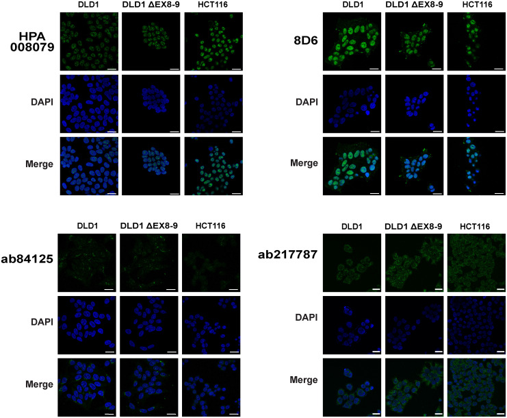
Fig 3. Immunofluorescence analysis of RNF43 antibodies.
DLD-1, DLD-1 ΔEX8-9, and HCT116 cells were cultured on glass slides and stained with HPA008079, ab84125, ab217787and 8D6 antibodies. DLD-1 ΔEX8-9 and HCT116 cells are not expected to reveal any staining, but in all cases signals are observed comparable with the wild-type control DLD1. DAPI was used to stain the nuclei. For the 8D6 antibody the DAPI-staining is coincidentally stronger in the DLD-1 ΔEX8-9 cells, giving the potential false impression that the green 8D6 signal is weaker in these cells compared with their wild-type controls. However, evaluation of multiple independent images shows that non-specific signals are of comparable intensity. Larger images, a higher intensity image for ab84125, and negative control test are shown in S2 Fig. Scale bar, 25um.

