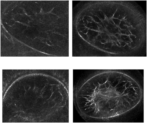
Figure 3.
Coronal breast images show SoftVue images on the right and prototype images on the left. A more fatty breast (top row) displays much better architectural detail, while the breast with more scattered central density (bottom row) is now clearly defined. Despite both breasts being scanned near the chest wall, the larger diameter of the SoftVue ring array than the prototype allowed clear boundary definition and much less near-field distortion from the breasts contacting the ring array (obscured upper and lower aspects of the top and bottom left images, respectively).

