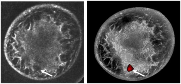
Figure 4.
SoftVue images show reflection-only reconstruction on left with an irregular but ill-defined 1.4cm invasive ductal carcinoma at the 6-7 o’clock position (arrows). The fusion image on the right used previously established thresholds to create a grayscale overlay which highlights the distribution of dense parenchyma. Moreover, the cancer is well seen when mass sound speed and attenuation thresholds were applied. Further work is needed to validate this highly encouraging initial fusion performance with other masses or suspicious foci.

