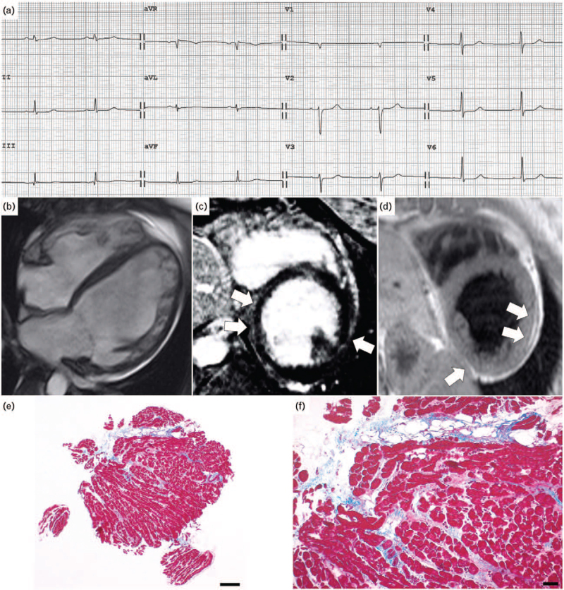
Fig. 1.
(a) ECG at admission showing sinus rhythm, low QRS voltages in limb leads, repolarization abnormalities in precordial leads and presence of premature ventricular beats; (b–d) contrast-enhanced CMR, (b) cine image with severe dilatation of LV and mild LV systolic dysfunction, mild dilatation of RV with preserved EF; (c) proton density weighting sequence showing fat infiltration of LV, involving mid segments of inferior and lateral walls (arrows); (d) postcontrast sequences showing a nonischemic subepicardial LGE in inferior and mid lateral walls (arrows); (e and f) endomyocardial biopsy with focal fibrofatty replacement of the myocardium [Heidenhain trichrome stain, scale bar 100 μm for (e) and 50 μm for (f)]. CMR, cardiac magnetic resonance; EF, ejection fraction; LV, left ventricular; RV, right ventricle.

