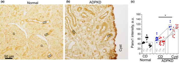
FIGURE 1.

Immunohistochemical staining in human autosomal dominant polycystic kidney disease (ADPKD) biopsy shows higher pannexin‐1 level in the cystic epithelium than in collecting ducts (CD). Representative pannexin‐1 staining in a normal kidney (a) and ADPKD patient with mutations in the PKD1 gene (b). G, glomerulus. (c) Summary graph of pannexin‐1 abundance. Normal kidneys (one female and two males) are represented by black symbols grouped per each of individuals; comparison CDs versus cysts in three ADPKD patient kidneys is shown as three pairs of values. Blue symbols denote data from the female patient with truncating frameshift mutation, red symbols—two male patients with nontruncating missense mutations. Boxes are mean ± SEM for each individual in the corresponding tissue. Columns are intensity mean ± SEM for each tissue type. *p < 0.05 calculated with unpaired or paired t‐test. N = 3 patients in each group.
