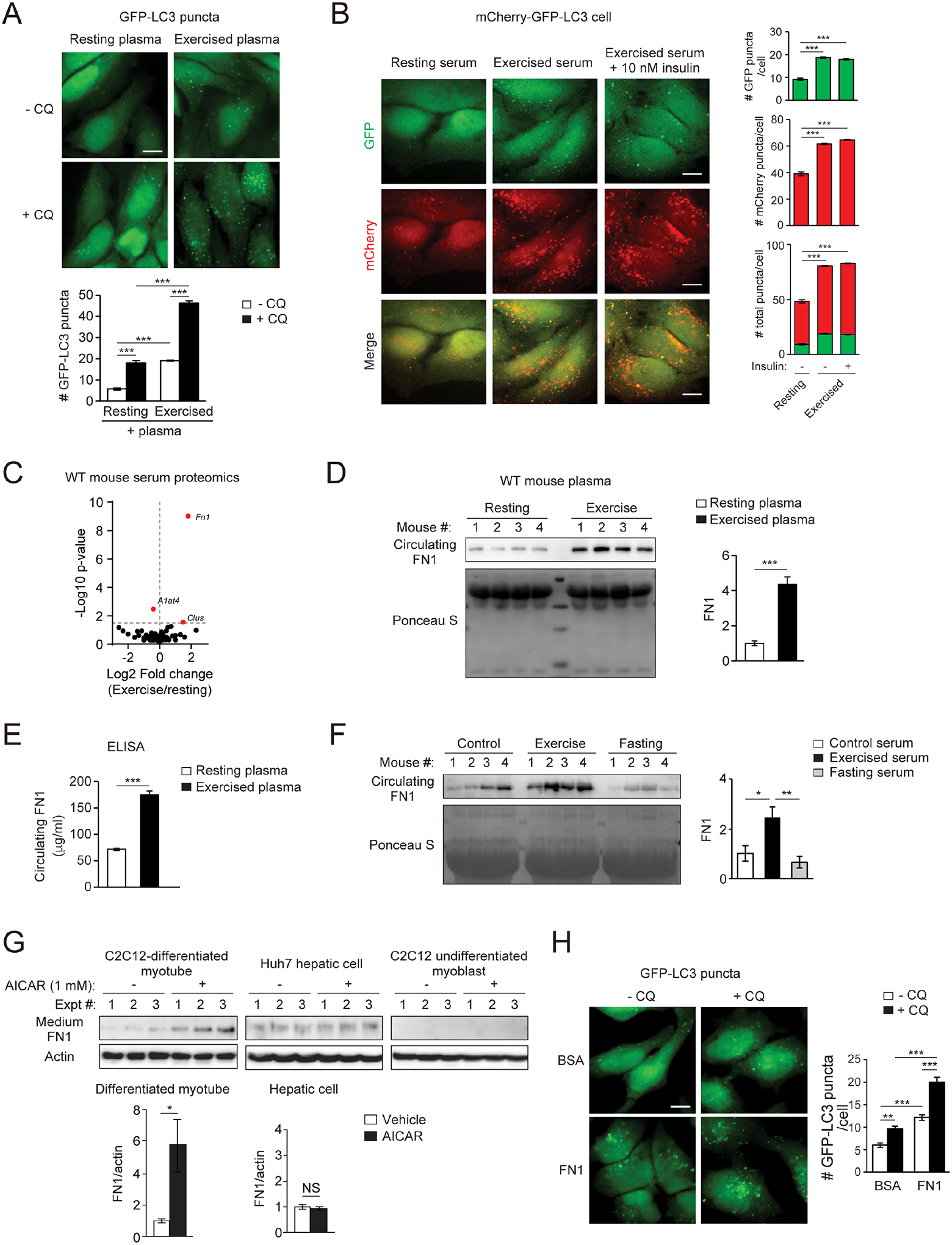
Figure 2. Identification of FN1 as an exercise-induced, muscle-secreted, autophagy-inducing factor.

(A) Representative images and quantification of autophagosomes (GFP-LC3 puncta) in HeLa cells stably expressing the GFP-LC3 reporter cultured for 3 h in medium containing 10% plasma from WT mice at rest or after 90-min treadmill running, with or without the lysosomal inhibitor chloroquine (CQ, 10 μM). Bar, 10 μm. N=3 mice. 50 cells per mouse were analyzed. (B) Representative images and quantification of autolysosomes (red) and autophagosomes (green) in HeLa cells stably expressing tandem mCherry-GFP-LC3 cultured for 3 h in medium containing 10% serum from WT mice at rest or after 90-min treadmill running with or without supplementation of 10 nM insulin. N=3 mice (30 cells/mouse serum treatment). Bar, 10 μm. (C) Volcano plot of mass spectrometry analysis on serum of WT mice at rest or after 90-min exercise. (D) WB of circulating plasma FN1 in WT mice at rest or after 90-min exercise. N=4. (E) ELISA of plasma FN1 in WT mice at rest or after 90-min exercise. N=10. (F) WB analysis of circulating FN1 in WT mice under fed and rested conditions, or after 90-min exercise or 24-h fasting. N=4. (G) WB analysis of FN1 in the conditioned medium of C2C12-differentiated myotubes, Huh7 hepatic cells, and C2C12 undifferentiated myoblasts treated with vehicle or 1 mM AICAR (AMPK activator) for 1 h. N=3. (H) Representative images and quantification of GFP-LC3 puncta in GFP-LC3 HeLa cells treated with or without FN1 or BSA (bovine serum albumin) (100 μg/ml) and the lysosomal inhibitor chloroquine (CQ) for 3 h. Bar, 10 μm. N=50 cells/condition. (A-B, F, H), one-way ANOVA with Tukey-Kramer test. (D-E, G), t-test. *, P<0.05; **, P<0.01; ***, P<0.001; NS, not significant.
