Figure 2.
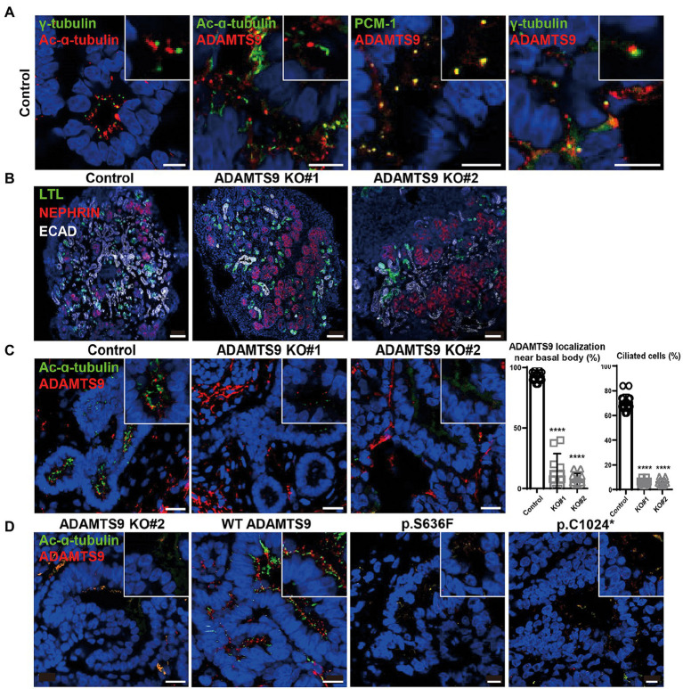
ADAMTS9 knockout kidney organoids demonstrate abnormal cilia, and ciliogenesis is not rescued by overexpressing ADAMTS9 variants. (A) ADAMTS9 is localized near the basal bodies of primary cilia and co-localized with PCM-1. The organoids were stained with anti-ADAMTS9, anti-Ac-α-tubulin, anti-PCM1, or anti-γ-tubulin antibodies. Scale bar, 10 μm. (B) Immunofluorescence staining of the kidney organoids displaying different structures, including the glomerulus (NEPHRIN), proximal tubules (LTL), and distal tubules (ECAD). Scale bar, 100 μm. (C) Loss or shortening of primary cilia was observed in ADAMTS9 knockout organoids. Percentage of ciliated tubular cells in kidney organoids and quantification of cilium length on the basis of Ac-α-tubulin staining are shown in the right panel. Data represent means ± standard deviation (SD). Scale bar, 10 μm. ****p < 0.0001. (D) Ciliogenesis was rescued by overexpressing wild-type but not by mutant forms of ADAMTS9, in ADAMTS9-ablated kidney organoids. Scale bar, 10 μm.

