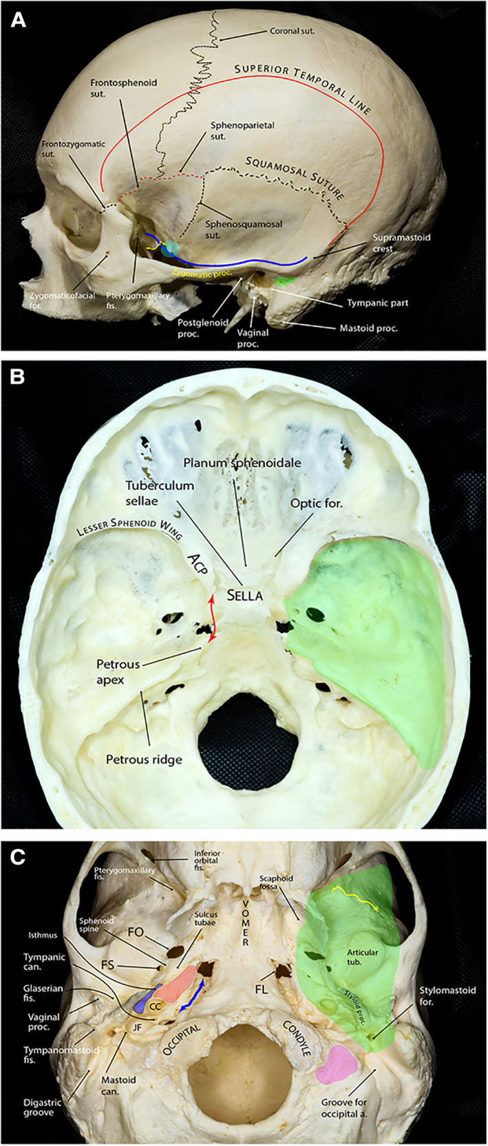
Figure 1.

Bony anatomy of the middle fossa. (A) Lateral view of skull showing relevant osseous landmarks for middle fossa surgery. Superior temporal line is the line of attachment of the temporalis muscle. The greater sphenoid wing and the temporal squama slope laterally and superiorly from the middle cranial fossa. The blue line shows the contour of the middle fossa floor projected on the lateral skull surface. The floor is almost at the level of the zygomatic arch. The floor ascends posteriorly and is approximately at the level of the supramastoid crest, which continues as the superior temporal line. At the level of the supramastoid crest, the middle fossa is separated from the tympanic cavity by the tegmen tympani. The suprameatal (aka McEwen) triangle (green area) lies just inferior to the supramastoid crest and posterosuperior to the external auditory canal and is the gateway to the mastoid antrum during mastoidectomy. Further anteriorly, the middle fossa floor is composed of the greater wing of the sphenoid that makes the roof of the infratemporalfossa. A prominent rough bony tubercle (sphenoid tubercle, cyan circle), which serves as one of the attachment points of the deep temporal fascia, appears at the lateral end of the infratemporal crest (yellow double arrow) to which the lateral pterygoid muscle is attached. (B) Endocranial view showing the middle fossa proper (green area) separated from the sellar and parasellar compartments by the petrous-clinoid line (red double arrow). The posterior boundary of the middle fossa is formed by the petrous ridge. (C) Exocranial view of the bony skull base. The green shaded area shows the approximate projection of the middle fossa floor on the exocranial surface. The bony part of the pharyngotympanic tube (blue) runs parallel and lateral to the carotid canal (red area) with its anterior end turning into the cartilaginous portion at the region of sulcus tubae opening into the nasopharynx. Yellow double arrow marks the infratemporal crest and the petroclival fissure is marked by the blue double arrow. Pink area shows the jugular process of the occipital bone. a., artery; ACP, anterior clinoid process; can., canaliculus; CC, carotid canal; fis., fissure; FL, foramen lacerum; FO, foramen ovale; FS, foramen spinosum; for., foramen; JF, jugular foramen; proc., process; sut., suture; tub., tubercle. (Copyright Ali Tayebi Meybodi. Used with permission.)
