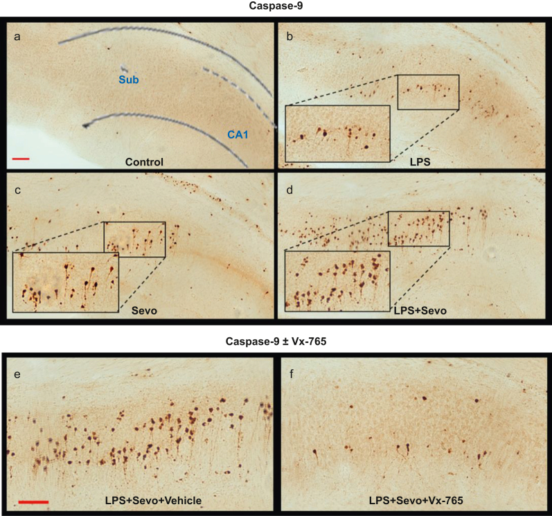
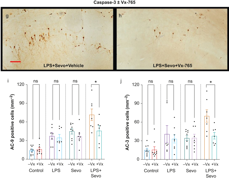
Fig 5.
Histomorphological analysis of activated caspase-9 (AC-9)-positive cells in subiculum and effects of Vx-765 pretreatment. (a–d) Representative images of AC-9 staining in subiculum. Images show AC-9 staining profile similar to that observed with AC-3. Scale bar is 200 μm. (e–h) Side-by-side comparison of representative images of AC-9 (e, f) and AC-3 (g, h) staining in subiculum of animals treated with LPS+sevoflurane, with vehicle vs Vx-765 pretreatment, respectively. Vx-765 pretreatment reduced the number of AC-9 (f) and AC-3 (h) -positive cells compared with vehicle (e and g, respectively) in combined LPS and sevoflurane treatment. Scale bar=200 μM. Bar graphs showing the effect of Vx-765 (+Vx) vs vehicle (10% DMSO, –Vx) pretreatment on subicular AC-9 (i) and AC-3 (j) cell counts. In addition to being non-apoptogenic to controls compared with vehicle, pretreatment with Vx-765 was able to prevent a significant number of AC-3-positive neurones from undergoing apoptosis in subiculum, but had no statistically significant effect when animals were challenged with LPS or sevoflurane alone. Data shown as mean (sem). Two-way anova + Sidak's post hoc, ∗P<0.05. anova, analysis of variance; LPS, lipopolysaccharide; sem, standard error of the mean; Sevo, sevoflurane.


