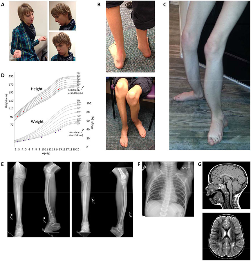
Figure 1. Physical exam findings and imaging abnormalities.
a. At age 15, mild facial dysmorphisms include triangular facies, anteverted nares, and micrognathia. b-c. Lower limbs with bilateral anterior tibial bowing, flat and pronated feet, and severe muscular atrophy (also at age 15 y). d. Growth curves demonstrating normal height (red dots), but weight (purple dots) that is low and decreasing in centile. Low weight was also present in the patients described by Leoyklang et al. [2007] and Döcker et al. [2014] (black dots). e. X-rays of the right (R) and left (L) lower legs (two views each) at age 9 y demonstrates anterior bowing of the tibias and fibulas with approximately 20° of angulation between the proximal and distal tibial segments. The diaphyses of the tibiae demonstrate hyperostosis. Diffuse osteopenia, non-weight-bearing pes cavus, and severe muscle atrophy are also seen. f. Chest X-ray at age 6 y demonstrating mild curvature of the mid-thoracic spine with convexity to the left (Cobb angle 15°). g. An MRI of the brain at age 3 y shows thinning of the splenium of the corpus callosum with evidence for cerebellar tonsillar ectopia (top, sagittal T1-weighted image) and gliosis of the bilateral posterior centra semiovale with associated dilation of the perivascular spaces (bottom, axial T2-weighted image).

