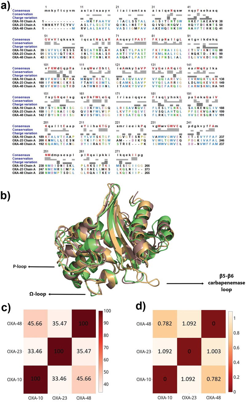
Figure 1.

a) Multiple sequence alignment of OXA-10 (PDB ID: 1FOF), OXA-23 (PDB ID: 4K0X), and OXA-48 (PDB ID: 3HBR). Consensus residues are colored and indicated in the upper case at the top row. The degree of conservation is presented in light grey. b) Aligned 3D structures of OXA-10 (light Orange), OXA-23 (pale green), and OXA-48 (dark salmon) highlighting the differences occurring especially on important loops surrounding active site cleft. c) Heatmap of sequential identities on a scale of 0 to 100, where 100 denotes the highest similarity. d) Heatmap of RMSD differences, where 0 denotes the highest similarity.
