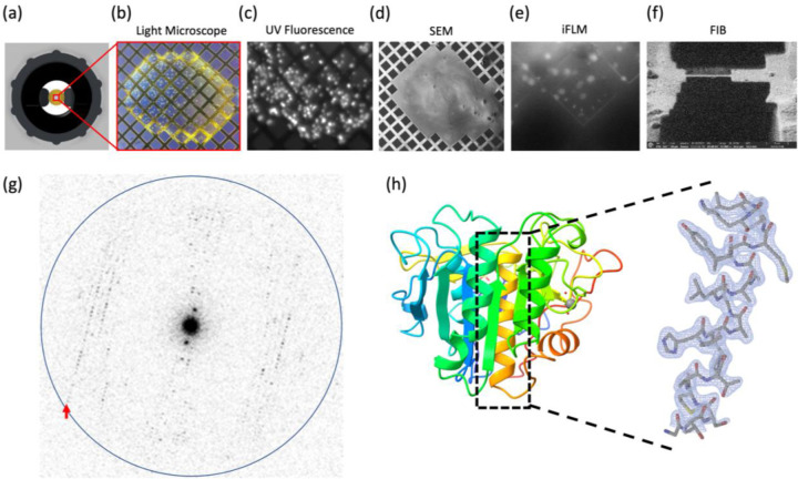
Figure 2. MicroED structure of suspended drop Proteinase K.
(a) The suspended drop viewed from the top and imaged by (b) Light microscopy or (c) UV. A frozen suspended drop specimen was loaded into the FIB/SEM and imaged normal to the grid surface by (d) SEM and (e) iFLM with the 385 nm LED to locate submerged crystals. (f) The targeted crystal site was milled into a 300 nm thick lamella. (g) Example of MicroED data acquired from the crystal lamella. The highest resolution reflections visible to 2.1 Å (red arrow). Resolution ring is shown at 2.0 Å (blue). (H) Cartoon representation of the Proteinase K colored by rainbow with blue N terminus and red C terminus. The 2mFo–DFc map of a selected alpha-helix is highlighted, which was contoured at 1.5 σ with a 2-Å carve.

