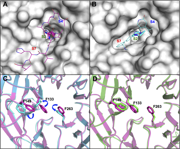
Figure 4.
(A) Surface representation of the WDR5 WIN site (PDB ID: 3EG6)14 when bound to MLL1 peptide (magenta carbon-capped lines and sticks (P2 residue)) with labeled S2, S4, and S7 binding regions. (B) Surface representation of the WDR5 WIN site (PDB ID: 4QL1)38 when bound to OICR-9429 (cyan carbon-capped lines and sticks (S2 binding moiety)) with labeled S2, S4, and S7 binding regions. (C) Overlay of MLL1 peptide (magenta) and OICR-9429 (cyan) bound WDR5 protein structures in cartoon representation. The side chains of WDR5 residues F133, F149, and F263 of both structures are represented as sticks to demonstrate conformational changes. (D) Overlay of MLL1 peptide (magenta) and compound 1 (green) bound WDR5 protein structures in cartoon representation. The side chains of WDR5 residues F133, F149, and F263 for both structures are represented as sticks to demonstrate retention of binding poses.

