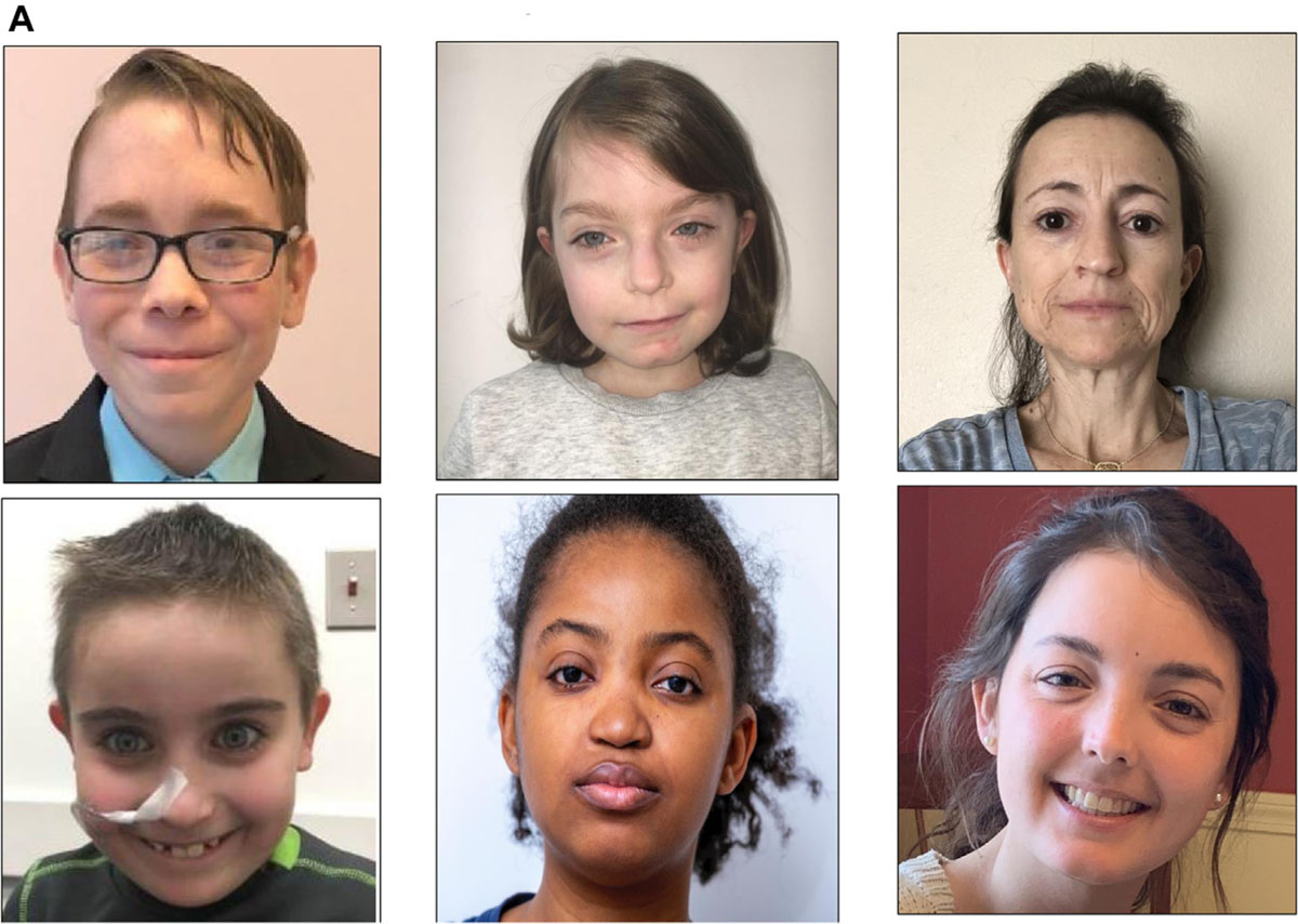
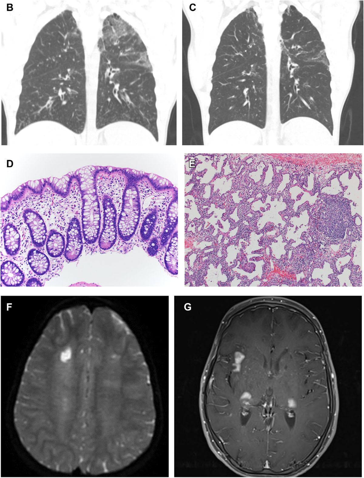
FIG 4.


Clinical images of STAT3 GOF patients. (A) Characteristic facial appearance includes round face, prominent forehead, cupped ears, and smooth philtrum. (B) Interstitial infiltrate demonstrates nodules in right upper lung, bronchiectasis, and airspace disease (patient 57). (C) Same patient as (B) after treatment with tofacitinib for 5 years showing resolution of ground-glass opacities and pulmonary nodules. (D) Rectosigmoid biopsy sample reveals mild eosinophilia in the lamina propria and chronic changes in the form of Paneth cell metaplasia (patient 61). (E) Lung biopsy of right lower lobe reveals nodular and linear septal inflammation, predominantly by lymphocytes and histiocytes, without interstitial fibrosis (patient 61) (hematoxylin and eosin, original magnification 100 ×). (F) Chronic ischemic lesion is in the white matter of the middle frontal gyrus and right semioval center, and extensive leukoencephalopathy is evident of probable microvascular origin (patient 152). (G) Subcortical and cortical enhancing lesions are in the presylvian regions, anterior temporal lobes, and pons (patient 110).
