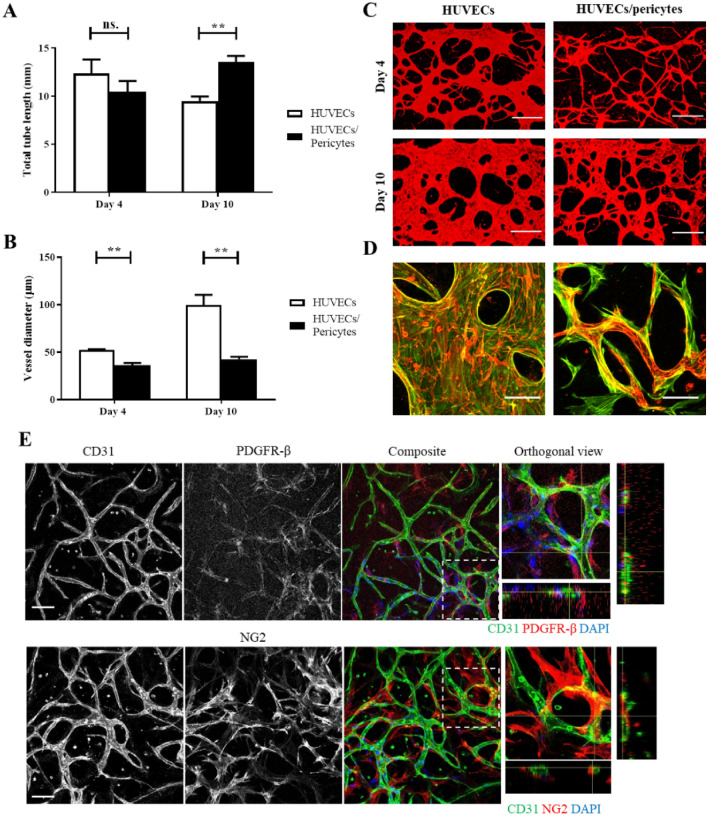
Figure 2.
The impact of pericytes on vessel length and diameter. (A) In direct co-cultures, pericytes had no significant impact on total tube length at day 4 (mean ± SEM, 12.3 ± 1.5 vs 10.4 ± 1.1 mm/field of view, HUVECs vs HUVEC/pericytes respectively), however, a significant increase in total tube length is seen by day 10 (mean ± SEM, 9.5 ± 0.5 vs 13.5 ± 0.6 mm/field of view, HUVECs vs HUVEC/pericytes respectively). (B) The addition of pericytes also led to a significant reduction in vessel diameter following both 4 and 10 days of culture. (C) Representative images of samples, z-projection images were generated using confocal images. Red, CD31. Scale bar: 300 µm. (D) Representative images showing HUVEC-pericyte interactions, z-projection images were generated using confocal images. Red, CD31. Green, F-actin. Scale bar: 100 µm. N = 3; n.s., non significant; **, p < 0.01. (E) Cells expressing the pericyte-specific markers PDGFR-β and NG2 (red) are clearly seen at the surface of the microvascular network. Corresponding orthogonal views indicate that these cells are often found wrapping around CD31 positive vessels. Scale bar: 100 µm.

