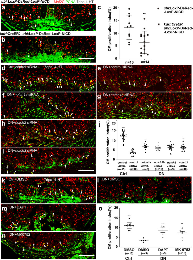
Fig. 4. Endothelium-specific expression of NICD or dn-xbrg1 decreases cardiomyocyte proliferation that is partially rescued by inhibition of Notch signaling.
a, b Immunostaining showing that Mef2C+ and PCNA+ proliferating cardiomyocytes of control (a) and endothelial NICD-overexpressing heart sections (b) at 7 dpa after 4-HT induction. c Statistics of panels (a) and (b) (data are the mean fold change ± s.e.m.; **p < 0.01, unpaired t-test). d–i Representative images of immunostaining showing that, compared with control siRNA treatment (d), PCNA+/Mef2C+ proliferating cardiomyocytes decreased at 7 dpa in DN-xBrg1 hearts (DN) treated with control siRNA (e), which were partially rescued by either notch1a (f), notch1b (g), notch2 (h), or notch3 (i) siRNA treatment in the presence of 4-HT. Scale bar, 100 μm. j Statistics of panels (d–i) (data are the mean ± s.e.m.; *p < 0.05; **p < 0.01; ***p < 0.005; one-way analysis of variance [F = 32.5] followed by Dunnett’s multiple comparison test). k–n Representative images of immunostaining at 7 dpa showing that, compared with DMSO treatment (k), PCNA+/Mef2C+ proliferating cardiomyocytes in DN mutant hearts decreased (l), which were partially rescued by either DAPT (m) or MK-0752 treatment (n) in the presence of 4-HT. Scale bar, 100 μm. o Statistics of panels (k–n) (data were the mean ± s.e.m.; ***p < 0.005; one-way analysis of variance [F = 22.4] followed by Dunnett’s multiple comparison test). n number shown here (c, j, o) indicated biological replicates.

