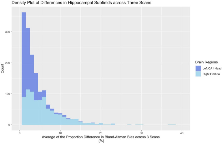
Fig. 1.
Bland–Altman plots of the average difference for volume estimation across subjects’ three MRI scans for the Left Cornu Ammonis (CA) 1 Head (dark blue) and Right Fimbria (light blue). The horizontal axis indicates the average difference in Bland–Altman “bias” (difference between subregional volume output for different scans, as a proportion of a region’s volume), while the vertical axis indicates the number of scans with a given value. Of note, the left CA1 Head has a low degree of mean bias (as a proportion of the region’s volumes; 0.106%), while the right Fimbria has a fair degree of mean bias (1.198%)

