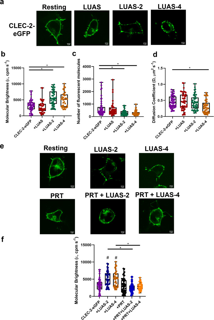
Fig. 2. Multivalent CLEC-2 nanobodies cause clustering of CLEC-2 and Syk plays a critical role.
a Representative confocal microscopy images showing membrane localisation of CLEC-2-eGFP under basal condition and treated with LUAS (10 nM), LUAS-2 (10 nM) and LUAS-4 (10 nM) for 60 min in transfected HEK293T cells (field of view = 52 × 52 μm) (scale bar = 5 μm). Box plots showing the effect of LUAS (10 nM) LUAS-2 (10 nM) and LUAS-4 (10 nM) on CLEC-2-eGFP b molecular brightness (ε, counts per molecule per second, cpm s−1), c number of fluorescent molecules within the confocal volume and d diffusion coefficient. FCS measurements were taken in 45–50 cells (n = 3 biologically independent experiments). e Representative confocal microscopy images showing membrane localisation of CLEC-2-eGFP resting and treated with LUAS-2 (10 nM), LUAS-4 (10 nM), PRT-060318 (10 μM), PRT (10 μM) + LUAS-2 (10 nM) and PRT (10 μM) + LUAS-4 (10 nM) in transfected HEK293T cells (field of view = 52 × 52 μm) (scale bar = 5 μm). Samples were pre-treated with PRT for 45 min prior to agonist addition for 60 min. f Box plot showing the effect of the treatments on the molecular brightness (cpm s−1) of CLEC-2-eGFP. FCS measurements were taken in 31–49 cells (n = 3 biologically independent experiments). For all box plots, centre lines represent the median; box limits indicate the 25th and 75th percentiles and whiskers extend to minimum and maximum points. Significance was measured with Kruskal-Wallis with Dunn’s post-hoc where P ≤ 0.05. In (f) # = significance compared to CLEC-2 alone (no ligand).

