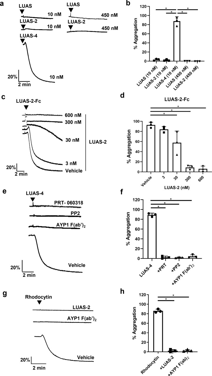
Fig. 3. Human platelets are activated by tetravalent but not divalent CLEC-2 nanobodies.
Platelet aggregation was monitored by light transmission aggregometry at 37 °C with constant stirring at 1200 rpm for 5–10 min. a Representative traces of platelet aggregation (2 × 10⁸ platelets/ml) following incubation with monovalent LUAS, divalent LUAS-2 and tetravalent LUAS-4 (10 nM). b Percentage of platelet aggregation induced by LUAS, LUAS-2 and LUAS-4 (n = 3 biologically independent experiments). c Representative traces of platelet aggregation induced by tetravalent LUAS-2-Fc (1.25 nM) in the absence (vehicle) and presence of increasing concentrations of divalent LUAS-2 (3, 30, 300, 600 nM). Platelets were preincubated for 5 min at 37 °C prior to stimulation. d Percentage of platelet aggregation induced by LUAS-2-Fc in the absence and presence of LUAS-2 (n = 3 biologically independent experiments). e Representative traces of platelet aggregation induced by LUAS-4 (10 nM) in absence (vehicle) and presence of PRT-060318 (1 μM), PP2 (20 μM) and AYP1 F(ab’)2 (66 nM). Platelets were preincubated with inhibitors for 5 min at 37 °C prior to stimulation. f Percentage of platelet aggregation induced by LUAS-4 with inhibitors (n = 3 biologically independent experiments). g Representative traces of platelet aggregation induced by rhodocytin (100 nM) in the absence (vehicle) and presence of LUAS-2 (10 nM), and AYP1 F(ab’)2 (66 nM). Platelets were preincubated with inhibitors for 5 min at 37 °C prior to stimulation. h Percentage of platelet aggregation induced by rhodocytin (100 nM) in the absence (vehicle) and presence of LUAS-2 (10 nM), and AYP1 F(ab’)2 (66 nM) (n = 3 biologically independent experiments). Significance was measured using one-way ANOVA with a Bonferroni post-hoc test where P ≤ 0.05. Data presented as mean ± SD.

