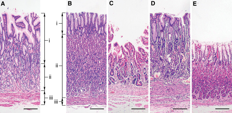
Figure 1.
Morphometric measurements from gastric antral (A) and corpus mucosa (B–E). Foveolar length (i) was measured from the upper border of the superficial epithelium to the base of the gastric pit. Glandular length (ii) was defined as the distance between the bottom of the foveolae and upper border of musculus mucosae. Musculus mucosae thickness (iii) was measured from the upper edge to the lower edge of the musculus mucosae. Total mucosal thickness was the sum of the above 3 values. Compared with non-atrophic corpus mucosa (B), foveolar length and musculus mucosae thickness were increased and the glandular length was decreased in atrophic corpus mucosa (C and D), while the total mucosal thickness was variable or no change (C and D). E showed total mucosal thickness thinning without histopathological atrophy (Hematoxylin and eosin staining, 40× magnification, bar = 200 μm).

