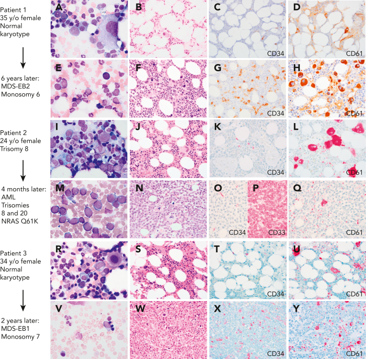
Figure 1.
Myeloid progression in GATA2 deficiency. (A-D) A 35-year-old (35 y/o) female with neutropenia, normal hemoglobin and platelet count, and very low monocytes, B cells, and NK cells count. (A) BM aspirate showed dysplastic MKs with minimal morphologic dysplasia in other lineages and no increase in blasts. (B) The biopsy was hypocellular with trilineage hematopoiesis, (C) low number of CD34+ cells, (D) few atypical MKs highlighted by CD61, and normal cytogenetics. (E-F) Six years later, she presented with severe anemia with 16% circulating blasts. (E)The marrow aspirate showed increased scattered blasts. (F) The marrow biopsy was hypercellular with (G) 10% CD34+ blasts, (H) increased CD61+ dysplastic MKs and increased reticulin fibrosis (not shown) indicative of MDS with excess blasts. Cytogenetics showed presence of monosomy 6. A germ line GATA2 mutation was subsequently identified (T354M). (I-L) A 24-year-old female with a prior diagnosis of aplastic anemia in adolescence presented with moderate pancytopenia and very low monocytes, B cells, and NK cells count. (I) The marrow aspirate and (J) BM biopsy showed normocellular marrow with trilineage hematopoiesis, (K) no increase in CD34 positive blasts, and (L) moderate dysmegakaryopoiesis. Cytogenetics revealed trisomy 8. Germ line GATA2 mutation was identified (L375V). (M-P) Four months later she presented with a platelet count of 10 × 103/μL. (M) The BM aspirate showed increased blasts. (N) The marrow biopsy was markedly hypercellular with (O) sheets of blasts that were negative for CD34 and (P) positive for CD33, and (Q) markedly decreased dysplastic MKs. Flow cytometry analysis of the marrow aspirate (not shown) identified 85% monoblasts that expressed CD56, CD64, CD36, and CD123 with minimal expression of CD14 indicative of acute monoblastic leukemia. Cytogenetics showed new trisomy 20 plus trisomy 8 in 65% of metaphases. (R-U) A 34-year-old female with mild pancytopenia, low monocytes, B cells, and NK cells count. (R)The marrow aspirate showed trilineage hematopoiesis with a subset of mononuclear MKs. (S) The BM biopsy was hypocellular for age with trilineage hematopoiesis with (T) no increase in CD34+ blasts, (U) mild megakaryocytic atypia, and trisomy 8. (V-Y) Two years later she presented with circulating blasts. (V) The marrow aspirate was paucicellular with scattered blasts. (W) The BM biopsy was markedly hypercellular with (X) 8% CD34+ myeloblasts confirmed by flow cytometry, and (Y) dysplastic megakaryopoiesis with microMKs. Cytogenetics showed new monosomy 7. Germ line GATA2 mutation was identified (N371K). Marrow aspirates were stained with Wright-Giemsa stain (1000×). BM biopsies were stained with hematoxylin and eosin or immunohistochemistry (IHC) as indicated (500×). Images were taken using an Olympus BX41 microscope equipped with a DP74 camera using Olympus cellSens software. EB1, excess blasts.

