
FIGURE 5.
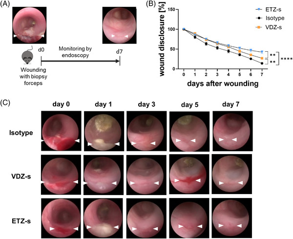
Delayed in vivo wound healing upon treatment with anti‐β7 integrin antibody. (A) Schematic depiction of experimental set‐up. On day 0, wounds were inflicted to the colon of C57BL/6J mice. From day 0 through 7, wound healing was monitored by endoscopy. (B) Quantitative analysis of relative wound diameters over time in mice treated either with anti‐β7 integrin antibody (n = 11), anti‐α4β7 integrin antibody (n = 8) or with isotype control (n = 10). (C) Representative endoscopic images at the indicated time points. **p < 0.01; ****p < 0.0001.