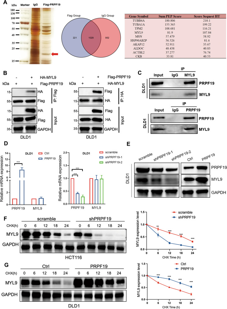
Fig. 3. PRPF19 stabilizes MYL9.
A DLD1 cells were treated with MG132 (20 μM) for 5 h, lysed and subjected to immunoprecipitation with anti-PRPF19 antibody or IgG using silver staining. The red arrow represents the detected bands. The potential interacting proteins of PRPF19 that possessed the highest score were listed (right panel). B The DLD1 cells were co-transfected with the indicated plasmids for 48 h, and cells were treated with MG132 for 5 h. The cell lysates were immunoprecipitated with antibody anti-Flag and immunoblotted with anti-HA (left panel); immunoprecipitated with antibody anti-HA and immunoblotted with anti-Flag (right panel). C DLD1 cells were treated with MG132 for 5 h before being collected. Cell lysates were immunoprecipitated with anti-IgG, anti-MYL9, or anti-PRPF19 followed by immunoblot. D, E The mRNA (D) and protein (E) expression level of MYL9 in the DLD1 stable cell lines. F, G The indicated stable cell lines with PRPF19 knockdown (scramble/shPRPF19) or overexpression (Ctrl/PRPF19) were treated with CHX, and lysed for WB analysis at the specific time points. The intensity of MYL9 expression for each time point was quantified with GAPDH as a normalizer. ***p < 0.001, **p < 0.01 based on Student’s t test.

