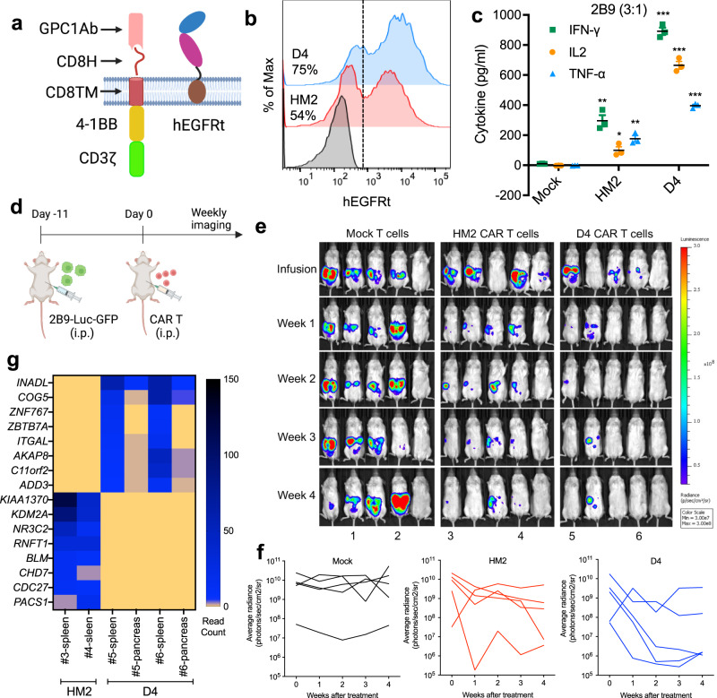
Fig. 2. GPC1-targeted CAR T cells eradicate tumors in the 2B9 intraperitoneal dissemination xenograft mouse model.
a Schematic of the GPC1-targeted CAR construct. The illustration was created with BioRender.com. b CARs expression on T cells were analyzed using flow cytometry by detection of hEGFRt expression. Data are representative of two independent experiments. c The secretion of IFN-γ, IL-2 and TNF-α in the CAR T and 2B9 co-cultured supernatant at E:T ratio of 3:1. n = 3 independent experiments. *p = 0.011, **p < 0.01, ***p < 0.001, two-tailed unpaired Student’s t test. d Experiment schematic (created with BioRender.com). 2B9 tumor-bearing NSG mice were treated with i.p. injection of 10 million mock T cells, HM2 CAR T cells and D4 CAR T cells on day 11 after tumor cell inoculation. n = 5 mice/group. e HM2 and D4 CAR T cells regressed established 2B9 xenografts in 4 of 5 mice in each group. f Tumor bioluminescence as photons per second in the Mock, HM2, and D4 CAR groups. g The heatmap of shared integrated genes in the D4 and HM2 CAR groups. The CAR T cells used in this figure were produced using the donor 1’s PBMCs. Values represent mean ± SEM. Source data and exact p values are provided in the Source data file.

