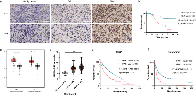
Fig. 1. The upregulated expression of IRAK1 is correlated with poor prognosis in glioma patients.
a Representative IHC images of IRAK1 protein in human glioma tissues and brain benign tumor tissues. 200×: scale bar = 100 μm; 400×: scale bar = 50 μm. b Kaplan-Meier survival plot for overall survival grouped by IRAK1 expression in 52 glioma patients (p = 0.0006). c IRAK1 mRNA expression profiling data from TCGA and GTEx databases analyzed by the GEPIA webserver (Non-tumor, n = 207; LGG, n = 518; GBM, n = 163; *p < 0.05). d IRAK1 expression analysis for non-tumor and glioma samples in Rembrandt database (****p < 0.0001). The correlation between IRAK1 and OS in glioma patients from TCGA (e) and Rembrandt (f) cohorts, with the hazard ratio (HR) and p values displayed.

