
Fig. 1.
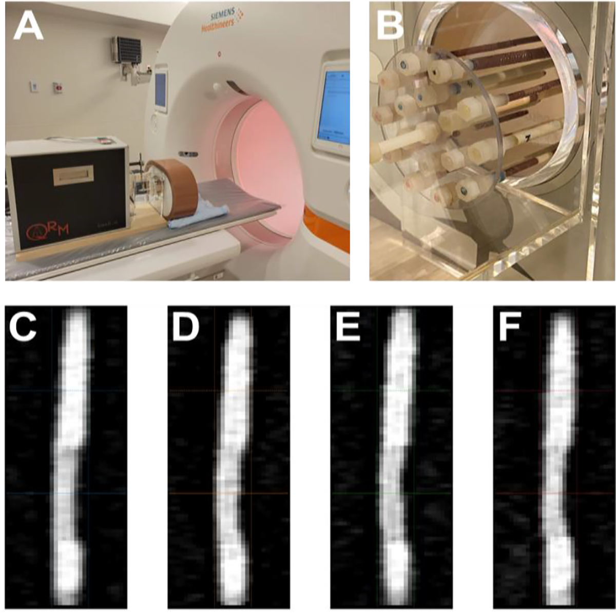
Experimental setup. (A) A cardiac motion phantom coupled with a 3D motion simulator was scanned with a dual-source photon-counting CT. It contained a variety of artificial coronary arteries containing stenoses of different materials and extents (B). 70 keV VMI images (C-F) illustrate a fibro-fat stenosis within the rod scanned at different heart rates (0, 60, 80, 100 bpm).