FIGURE 2.

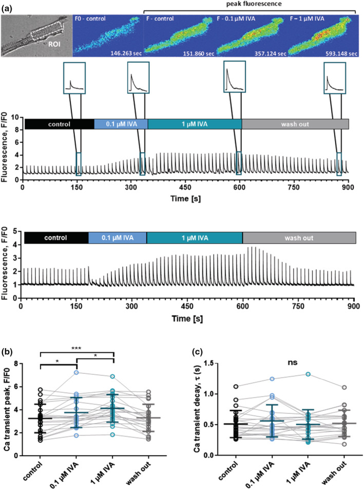
Acute effects of ivabradine (IVA) on intracellular Ca transients of ventricular cardiomyocytes derived from DMDmdx rats. The Ca transients were electrically induced at a frequency of 0.1 Hz. (a) Top panel: Representative transmitted light image (top left), and image series of false color Fluo‐4 fluorescence during the course of a typical experiment as shown below. xyt image series was acquired with a sampling rate of 90 ms per frame, and emitted Fluo‐4 fluorescence was spatially averaged from a region of interest (ROI) drawn in the central region of a cardiomyocyte. Middle and lower panel: Two original trace examples of intracellular Ca measurements. The blue color in the bars indicates the time periods of external ivabradine application. The insets on top show representative single Ca transients at an enlarged time scale under control conditions, in the presence of 0.1 and 1 μM ivabradine, as well as after washout of the drug. (b) Comparison of steady‐state Ca transient amplitudes under control condition, and in the presence of two different concentrations of ivabradine, as well as after washout of the drug. The data points connected with solid lines originate from individual ventricular cardiomyocyte experiments, respectively. The cells (n = 22) were derived from four independent preparations (4 DMDmdx rats). Data were also expressed as means ± SD. Repeated measures ANOVA revealed a significant difference between the experimental conditions (p < 0.001). *p < 0.05; ***p < 0.001 (Tukey's post hoc analysis). (c) Comparison of Ca transient decay kinetics under control conditions, and in the presence of two different ivabradine (IVA) concentrations, as well as after drug washout. The data points connected with solid lines originate from individual ventricular myocyte experiments, respectively. A single exponential function was fit to the decaying phase of the Ca transient in order to derive time constants (τ‐values). Ivabradine did not significantly affect Ca transient decay kinetics (ns, not significant; p = 0.33, Friedman test). Control experiments on ventricular cardiomyocytes isolated from a 9‐month‐old wild‐type Sprague Dawley rat revealed a significantly increased Ca transient peak (F/F0) in the presence of 1 μM ivabradine (3.6 ± 0.4 vs. 3.1 ± 0.4 in the absence of the drug; n = 11 cells; p < 0.01, Wilcoxon matched‐pairs signed rank test). Ca transient decay (τ‐value) was not affected by ivabradine (0.25 ± 0.06 s vs. 0.26 ± 0.04 s without drug; p = 0.83).
