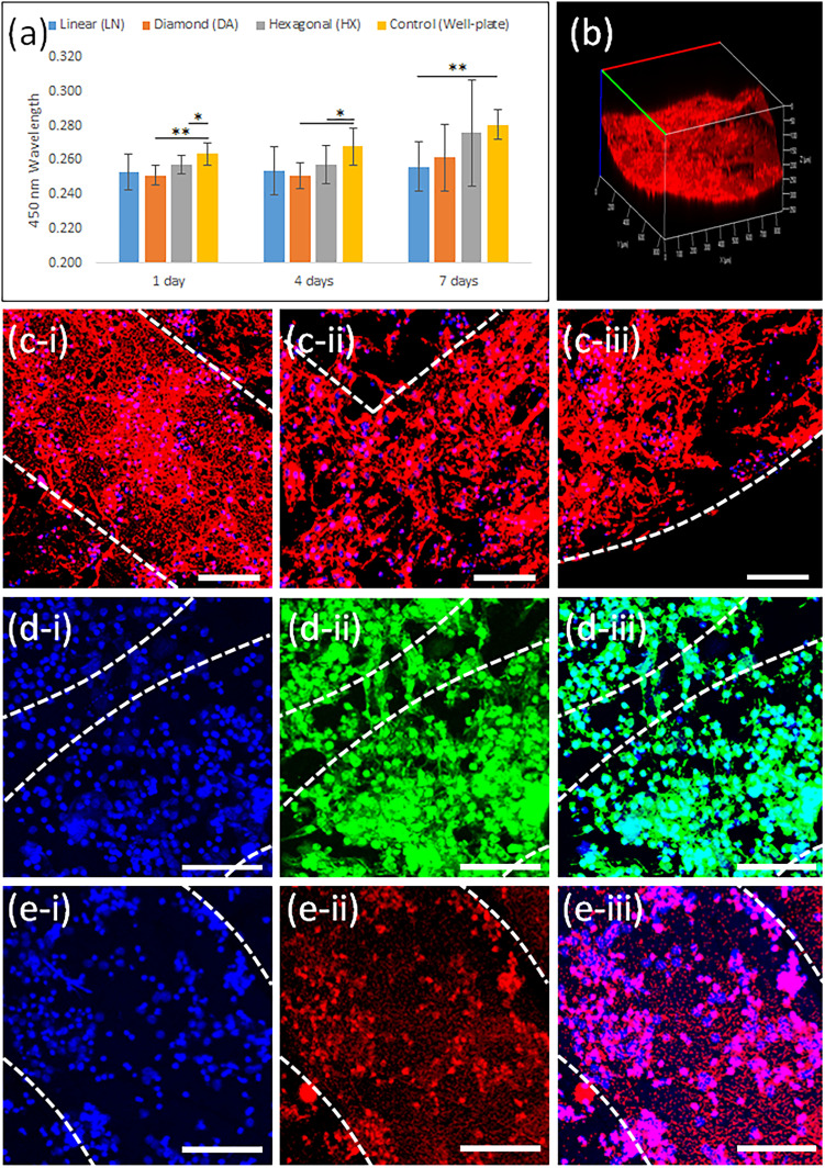
Figure 6.
(a) The proliferation of hiPSC-CMs on the 4D constructs compared to a 48 well-plate after 1, 4, and 7 days. Data are the mean ± standard deviation; n=9, *p<0.05 and **p<0.01. (b) A confocal microscope image of evenly distributed hiPSC-CMs on the 4D construct after 4D shape transformation using F-actin staining. Confocal microscope images of F-actin stained hiPSC-CMs on the 4D constructs with (c-i) LN, (c-ii) DA, and (c-iii) HX printing patterns after 1 week of culture. Scale bars, 100 µm. Confocal microscope images of α-actinin stained hiPSC-CMs on the HX 4D constructs after 1 week of culture with (d-i) nucleus, (d-ii) cardiac-specific protein, and (d-iii) combined. Confocal microscope images of cTnI stained hiPSC-SMs on the HX 4D constructs after 1 week of culture with (e-i) nucleus, (e-ii) cardiac-specific protein, and (e-iii) combined. Scale bars, 100 µm.

