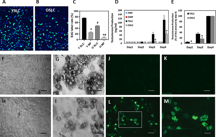
Figure 5.
Effect of aging on the proliferation and differentiation of CD51+ SLCs and CD51++ macrophages. (A, B) Young and old CD51+ SLCs were labeled with EdU for 6 hours in day 3 of the culture. (C) The rates of EdU+ CD51+ SLCs and CD51++ macrophages were quantified. (D) Testosterone production of CD51+ SLC and CD51++ macrophages after induction of differentiation. (E) Testosterone production by differentiated CD51+ SLCs on days 2 and 3 was expressed as percentage of day-4 cells. (F–I) Expression of HSD3B activity by CD51++ macrophages (F, H) or CD51+ SLC (G, I) 4 days after induction of differentiation. (F, G) Young cells. (H, I) old cells. (J–M) Expression of CYP17A1 proteins by young CD51+ SLC after 4 days in culture. (J) CD51+ SLC without primary CYP17A1 antibody. (K) CD51+ SLC before differentiation. (L, M) CD51+ SLC 4 days after induction of differentiation. ND: not detected. The data are expressed as mean ± SEM of three individual experiments. *,#Significantly different from age-matched YSLC or OSLC controls (*) or from same cell type from young animals (#) at P < 0.05 respectively. Black scale bars: 50µm in length. White scale bars: 20µm in length.

