Fig. 1.
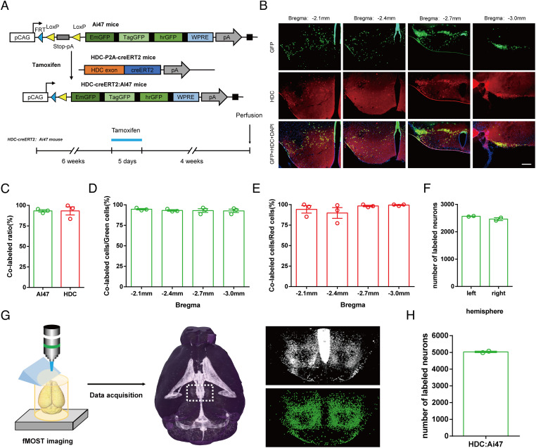
Whole-brain histaminergic neurons in the TMN of HDC-CreERT2:Ai47 mice. (A) Generation of HDC-CreERT2:Ai47 transgenic mice. Breeding Ai47 mice with HDC-CreERT2 mice yielded HDC-CreERT2:Ai47 mice, in which GFP is expressed specifically in HDC-positive neurons after injection of tamoxifen (100 mg/kg, i.p., 5 d). (B) Representative immunohistochemistry of expression specificity and penetrance in HDC-CreERT2:Ai47 mice on coronal sections with different distances from bregma. (Scale bars: 200 μm.) (C) Colocalization analysis of GFP and HDC in all sections of the posterior hypothalamus from 3 HDC-CreERT2:Ai47 mice. (D and E) Expression specificity (D) and penetrance (E) of coronal sections with different distances from bregma in HDC-CreERT2:Ai47 mice. (F) Count numbers of histaminergic neurons in the posterior hypothalamus of 2 HDC-CreERT2:Ai47 mouse bilateral hemispheres. (G) Main steps of data generation, acquisition, and processing in fMOST imaging. Raw data were analyzed by NeuroGPS system and manual correction. (H) Total numbers of labeled histaminergic neurons in the posterior hypothalamus of 2 HDC-CreERT2:Ai47 mice.

