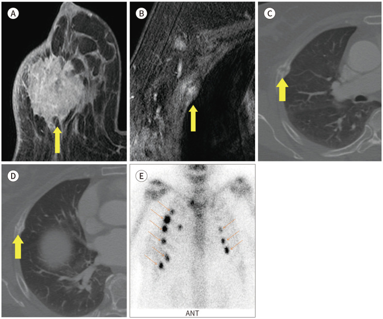
Fig. 20. 73-year-old female with IDC in right breast and rib fracture mimicking bone metastasis.
A. CE T1-weighted-MRI shows a 4.2 cm extent IDC in the right breast with skin invasion (arrow).
B. CE T1-weighted-MRI shows a focal enhancing lesion in right rib, which is suggestive of bone metastasis on the MRI (arrow).
C, D. Axial CE CT image also shows multiple rib fractures with callus formation (arrows).
E. Bone scan also shows multiple rib fractures in both sides (arrows).
CE = contrast-enhanced, IDC = invasive ductal carcinoma

