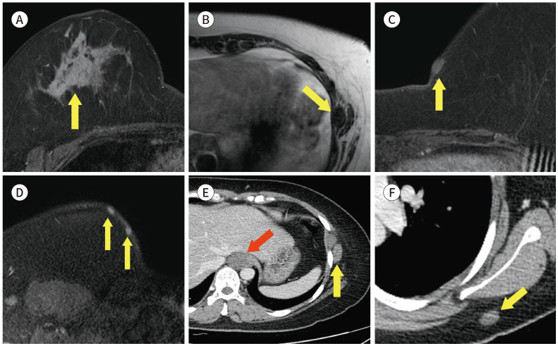
Fig. 22. 35-year-old female with IDC in right breast with history of neurofibromatosis.
A. CE T1-weighted-MRI shows a 7 cm in size IDC in the right breast (arrow).
B. T1-weighted-MRI shows a 1.6 cm in size nodule in the left lateral chest wall (arrow).
C, D. CE T1-weighted-MRI shows several small nodules in the subcutaneous layer (arrows).
E, F. In the CE axial CT, there is a 2.4 cm in size hardly-enhancing nodule in the paraaortic area (red arrow) and small nodules in the soft tissue (yellow arrows).
CE = contrast-enhanced, IDC = invasive ductal carcinoma

