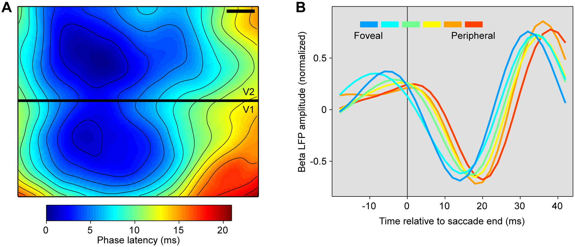
Figure 6. Evidence for waves in visual cortex traveling from stimulus location or from fovea.

(A) Map of phase latencies, of voltage-sensitive dye signal, at the border between awake macaque areas V1 and V2 (black bar indicates 1 mm on cortical surface). A visual stimulus had been presented at a position in visual space represented approximately at the locations with minimal phase latency. After stimulus presentation, a wave propagates over both V1 and V2, with phase latencies increasing systematically with distance from the stimulus’ representation. Adapted and modified from Muller et al.46.
(B) Amplitude of beta-filtered LFP as a function of time relative to the end of a saccade. Each line represents one of six recording sites in awake macaque area V4, representing visual field locations ranging from fovea to periphery, as indicated by the color legend. Adapted and modified from Zanos et al.47.
