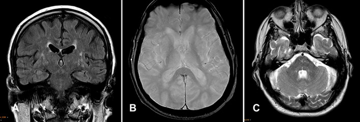
Fig 1.

The coronal T2 FLAIR sequence (A) shows widening of perivascular spaces and white matter hyperintensities in the white matter of centrum semiovale and basal ganglia suggestive of cerebral SVD. Axial gradient echo image at the level of the basal ganglia (B) demonstrates a normal signal without signal dropout, and the axial T2‐weighted image (C) at the level of the pons, middle cerebellar peduncles, and cerebellar hemispheres shows patchy non‐specific foci of high T2 signal in the pons.
