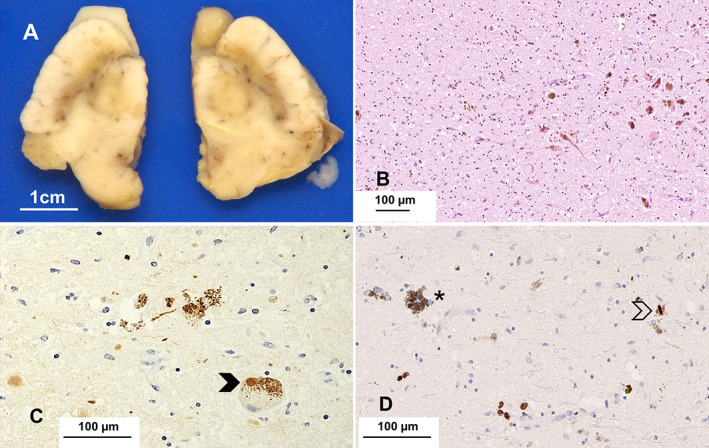
Fig 2.

The substantia nigra is pale bilaterally for the age of this patient. Microscopic examination reveals considerable loss of pigmented neurons and pigment incontinence (B, HE – scale bar) and Lewy bodies (arrow) and threads (C, α‐syn); the immunoreaction for Ki‐67 highlights neoplastic cells infiltrating the nigra (asterisk: pigmented neuron); a mitosis is present (arrow) (D, immunoperoxidase). Scale bars: 1 cm (A), 100 μm (B, C, D).
